Friday, January 13, 2012

Poor Leachey!! :( (leech lab)

Just to point this out, yes I did name my virtual leech Leachey!

If you would like to try this great lab please go to POOR LEECH!

In the lab, you first begin learning about the leech, or actually you start off by by using the leech tongs and try to grab the little parasite. You put the leech in 20% ethanol to numb it or in this case, knock it the heck out so it does not feel it's untimely death! Once you grab the leech from the ethanol, you place poor Leachey in the dissection tray. Poor Leachey, I had trouble pinning him down to the tray. I couldn't figure which side of him was his tail or his head....It looked the same! The big end is actually not the head....its the tail. So in the computer stimulation I was poking the poor leech's tail and not his head like 45 times before I FINALLY got the idea.
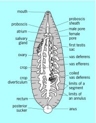


As I continued, I grabbed the scissors and managed to cut through the first layer of the leech (due to the angle of the scissors) and removed the inner tissue and guts of the leech and placed it under the disection microscope. I continued to romve certain parts of the tissue with the foreps into the light of the dissection microscope. If i did this in real life it seemed like the inside of the leech would seem much more well put then the stimulation gave. As you continued looking at the tissue and such of the leech with the microscope you press the tissue with a Oscilloscope which shows the voltage which can pass through the designated tissue and we can see which tissue responds to it and other stimuli such as a feather or a metal probe. YOu can see the reaction made with the Oscilloscope and the microscope and identify the tissue (ex: tissue t or s) It was difficult to identify the tissues as the would not respond to anything unless you found the exact spot the tissue responds too.
What I learned:
I learned that (as in the leech) has different muscles that react to different stimuli (in this case a feather, nothing, and electrical shock) and create different functions for each muscle to use. I also learned the leeches are parasites and can cause you to become sick due to the bacteria they carry in their mouth.

equipment:

                       Feather: Used to give the leech skin a very gentle touch stimulation. It really doesn't need to be a feather, it could be q-tips or something. Cost: free.

Probe: A blunt metal rod attached to a wooden handle useful for lifting, pushing, pressing, moving of specimen. Here you use it to lift tissue, and to push the skin as a stimulus. Typical price: $1.00 ~ 10.00
Forceps: Fine forceps for very fine manipulations. The very fine ones are known as Dumont #5 forceps, with tip size of about 0.1 mm X 0.06 mm or smaller. Typical price: $15.00 ~ 45.00

Scissors: Good dissecting angled scissors used here to cut open the body wall. Teaching scissors are cheaper, but some ultra-fine dissecting scissors could cost upward of $400, and you better not drop that, because once you drop it, chances are, it's ruined. Typical price: $15.00 ~ 60.00

Pins: Stainless steel dissecting pins for pinning tissue to a dissecting dish or board. You can drop these and not worry about it. $1.00

Scalpel: For microsurgery, disposable scalpel blades are better and much more economical than the fixed blade scalpel which needs to be sharpened periodically. Blade: $0.50 Handle: $10.00 Used here to cut all kinds of things.

Dissection Tray: A tray half-filled with hard wax so that you can stick pins into it to stabilize specimen for dissection.

Leech Tank: Leeches are kept in pond-water (you can actually buy an instant pond-water mix to add to tap water.) If kept in a refrigerator, they can stay happy in it for weeks at a time without feeding.

20% Ethanol: Used to anesthetize the leech. Besides being more humane, it has the added benefit that it stops them from moving, making it easier to pin down the leech.

Leech Tongs: These are basically gross anatomy forceps with blunt tips so that you will not harm the leech as you pick it up. Maybe about $ 10.00

Dissection Microscope: These are binocular microscopes specifically designed for dissection and other micromanipulations. Essentially, it's a high quality high power magnifying glass. The price varies on quality and if you've looked through binoculars of different quality, you can appreciate what a difference good optic makes. On a good one, you can clearly see individual cells in a leech's nervous system. Cost about $1,000.00 ~ $7,000.00

Micromanipulator: A device used to position items with sub-micrometer precision in three dimensions. Here we mount our electrode on it to guide it accurately to a neuron. For work on a leech, a mechanical manipulator would suffice which is about $700.00. More accurate hydraulic or electronic ones may cost up to $10,000.00

Oscilloscope: Basically a sophisticated voltmeter. What you see on the screen is a real time display of voltage (vertical) plotted against time (horizontal). Useful because voltmeters can't track rapidly changing voltages, and even if they could, you couldn't read anything. Cost $2,000.00 and up.

Leech: Medicinal leeches are about $15.00 each. When fully extended, they can reach 15 to 20 cm long. When fully contracted, diameter is roughly 1 ~ 2 cm.

Thursday, January 5, 2012

nervous system

The nervous system is the "master control" for controlling and communicating with your body.


Sensory input (Afferent)-Stimuli both and inside outside of the body
Integration-From sensory input
Motor output (Efferent)- Response to stimuli

The central nervous  system is the the brain and spinal cord are the integration and command  center


As the nerves spread down it spreads out like your fingers. another good example is your blood veins to capillaries. They just get smaller and smaller. They lead to neurons and axons. It is the areas where you receive amount of stimuli that sends messages up the nerve through your spinal cord to your brain and the action is received. Further action is taken from there as the brain sends a corresponding message 


Such as if a athlete strained the muscle and caused deep pain for the body. The body does not perceive the pain at first due to the amount of adrenaline the body has coursing through the system. yet as the athlete cools down and relaxes after a hard workout or such, the axons and neurons from the nervous system can now pick up on minuscule pain points in the muscle. depending on the amount of pain the point can give off the athlete can be sore, as in the muscles feel fatigued and stretched, to a harsh pain that has the athlete, limping and hobbling. That would be the deep pain the muscle feels, but the body perceives it through the messages the brain receives from the neurons and axons.


Another form of deep pain can hardly be perceived in the body. Say for instance a person is thrown from a horse and suffers a broken spine or neck and survives the incident. He/she cannot feel the pain that the body is suffering because the connection to the brain via the spinal cord is severed. The nerves, axons, and neurons can be sending the messages, that the body is hurt and needs medical attention.


For the nervous system to actually work, it depends on the message system it has set up. Axon to neuron, to the spinal cord to the brain where the brain then sends the appropriate action to the message it has received.


random fact: nuns have larger neurons then regular people

Tuesday, December 13, 2011

eating lab

This lab was excellent. In a procedure, Leeann had Seth and I attach some muscel reactive  adhesive sticky things (I honestly do not remember what they are called). These little stickies were attached to two points on our jaw and another to a different point on our body (both Seth and I opted for a pulse point). Seth went first and ate a different variety of foods and drank a large gatorade. With the data Seth gave us, it seems like he does not use a lot of energy to chew anything. He averaged around a .788 energy even used to eat his items.

I on the other hand seemed to use a a extream amount of energy for little things. an average of .865. I did have a jaw ache though so that may have factored into it. Out of the both of us, we both averaged a .827. We learned that it varies on the individual on how much jaw power you need to eat. We also learned that peanut butter makes me act like a dog with peanut butter stuck to the top of its mouth. Apparently it was amusing.

Monday, November 14, 2011

Bones bones---and bone breaks --\--

Well I should know a lot about bones. I think my family holds the record of the most broken bones in a life time.

My father has managed to crack his skull, break his ankles, and have a compound fracture all in 25 shorts years of his early life. Just those years! I have managed to disable every single bone in my body, break my fingers and half of my toes. My mom has busted her own ankle. My brother has also broken his nose. Now tell me that is not a record?!

Most of our fractures were hairline.
such as this. It was barley noticeable in the X-rays and not much could be done. It was excruciating pain.
Now the next fracture is what I had with my big and pinky toe on my left foot.
yet I have also had a stress fracture on my foot as well. I had jumped at the same time as a soccer player and my toes collided with his cleats. It was enough stress to bend my toe back and create and compound fracture.

Now with my pinky...it was a complete fracture. When I mean complete, I mean like the bone snapped and now my poor pinky is crooked. The thinner bone snapped , and was moved to the side. I never went to the doctors to fix it. I was after all in the middle of a great freshman season of basketball and I was sure I can finish the season with some tape and gauze protecting my hand. It made it worse really.

just like this. The bone continued to repair itself and the calcium build up has left my pinky crooked and a slight calcium deposit giving my joint a rough rounded appearance.
Now my dad's injuries were more...extensive and slightly funny at his own expense. (sorry dad!)

This is probably the most common injury both me and my father have sustained.
you guessed it. Skull fracture. See both me and my father have sustained blunt force trauma to the head multiple times. IE basketball accident, football accident, just falling on cememnt. Everything to manage a head injury, we have probably done it.

Examples of reciving said skull fractures through blunt force trauma....
case number one: This shall be my story. It was during a basketball season my freshman year. Simple request from my coach ("Get me the *bleep* ball Wolf! <---- that was my given name in the season)  and I was chasing down a 200 lbs. girl from Karval. Long story short: I caught her, my own teammate used me and her as a stop wall i crack my head on the only patch of cement in the gym with ove 300lbs on me.

case 2: now imagine that with my dad. Full football gear without helment messing around with friends. Yea not so pretty.

Now on to the fun part.

Compound fractures. Thank goodness but unforunatley my father has. It was at his elbow joint. His elbow broke falling out of a cherry tree. Originally he and his sister thought it was a stick that was sticking out of his arm but it actually turned out to be a long bone. He would not have survived if his grandmother had not inserted her finger into the gaping wound and cut off the blood flow from his artery.

just at the elbow.

Now on to actual bones. It might explain more about fractures if I do this right.

THere is several different shapes of bones.
long, short, flat, and irregular.

Indside the bone is where a lot of the body magic happens.
We have compact bone which has no poruous lok about it at all. It is solid bone. Spongey bone is not yet mature bone that has tiny holes in it making it look like sponge. This is a classic example of the long bone in our bodies.

Thursday, October 27, 2011

integumentary system

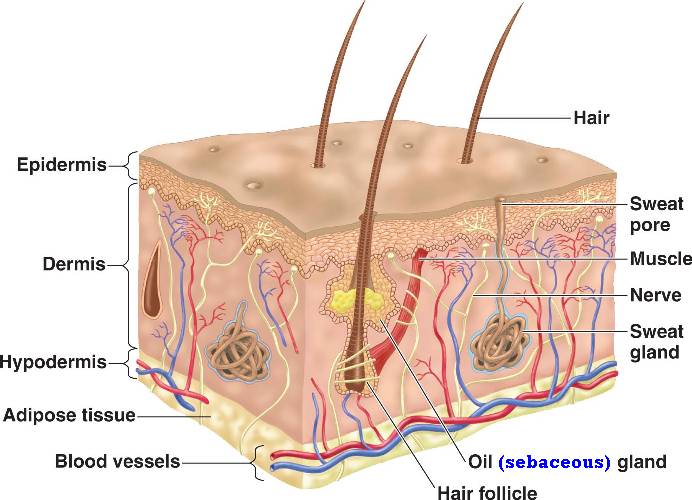
The integumentary system in our bodies is...wait for it.....our skin!

Yes the skin on your body is the integumentary system. It protects our bodies from the outside world. The air, the bugs in the world, and even the sun.

The epidermis is the top layer. It is completely dead skin. The skin is like a onion. It has many layers. Each layer rises and the dead skin is the top layer that is rubbed off. Interesting/ disturbing fact that I learned was you would have to wash your bed sheets everyday to get rid of the skin flakes that are rubbed off in your sleep.

Now the major layer in your integumentary system is the dermis. It holds major functions such as the the sweat glands and nerve endings. Also within this layer, are the capillaries. Another interesting fact is, when you lay in the sun (if fair skin you can see it the best) and you feel your skin heat up, it is your skin releasing the heat. The capillaries have expanded, making your skin seem red and is releasing the heat that is stored in your skin. It is acting as a refrigerator.

Now onto the fun part of this post. Skin diseases ....

Not all of them are dangerous but there are some that are life threatening. Such as... the bubonic plague (more commonly known as the black plague).
Which is caused by these little buggers
 Which ride on these guys
Who stow away on boats and the flea jumps to people. Thus transferring the bacterial infection which caused painful swelling of the lymph nodes in your system and making them burst open from the overload of puss and blood. There are three types of the "plague". Not all of them have the swelling though so be careful.

Friday, October 14, 2011

Tissue engineering review

Tissue Engineering

Summary:


The new development of bio genetic growing of tissue cells is now upon us. With the help of genetically altered rats, NASA, and the fore fathers of cell/tissue growth (Dr. Joseph Vacanti and Bob Langer). Tissue engineering is the basis for helping people who are needing to replace a organ or other features such as cardiac tissue, veins, cartilage, and skin. Thus it makes it easier for people who have a difficult time receiving help for a transplant. Way easier. The transplant actually works as well, even though it can be grown from a live specimen or even in NASA no-space container.


Weird example:
Photo  of heart valve


See! It's one of the genetically altered rats that is growing a ear for a person.... please don't shudder like I did.


Opinion:


*shudder fit*


Ok that is out of the way. See this is a new advancement from the past ( this all started in 1987). Now people don't have to be on the airing list for long anymore and people can also be treated for life threading illnesses like bone marrow transplants and cartilage transplants.

Tuesday, October 11, 2011

Histology

Histology: The study of the form of structures seen under the microscope. Also called microscopic anatomy, as opposed to gross anatomy which involves structures that can be observed with the naked eye. Traditionally, both gross anatomy and histology (microscopic anatomy) have been studied in the first year of medical school in the U.S. The word "anatomy" comes from the Greek ana- meaning up or through + tome meaning a cutting. Anatomy was once a "cutting up" because the structure of the body was originally learned through dissecting it, cutting it up. The word "histology" came from the Greek "histo-" meaning tissue + "logos", treatise. Histology was a treatise about the tissues of the body and the cells thereof.
Histology

I have studied cranial, epithelial,skeletal, and bone slides. Each were very different. In the lab we did, we were tought how to use a microscope and studied the afore mentioned slides under a 10x focus and then a 40x focus. we were told to draw and identify each slide. We did this and I shall post a good quality picture soon.

Epithelial:
  This is just one example of epithelial tissue. It can also com in squamous, columnar, and pseudo-.

Cranial: is well from your head. It can also come in cuboidal, squamous, columnar, and pseudo-. There are no real good pictures I could find. Sorry.

Bone:
Here are two really good pictures (one of which I stared at under a microscope which was extreamley amazing). This is bone tissue. You can tell it is by the circular pattern in the second picture.

 This will also fall under the skeletal tissue catagory.

Anyways. I was also doing a lab with Cassy, and Jena. We learned how to use the microscope exceedingly well. Focusing was a cinch for us and we all took turns drawing what we saw. Unfortunately Jenna threw our drawings away because they could not be transferred to the computer well. This was a interesting lab, we had to guess at what we saw. Almost like a test to ourselves. And we had fun for this lab which made it worthwhile. Though we could not spell or pronounce any names we learned each slide slowly and it was worthwhile. I would like to thank Mr. Ludwig for that.

please forgive the spelling mistakes spell check broke on me