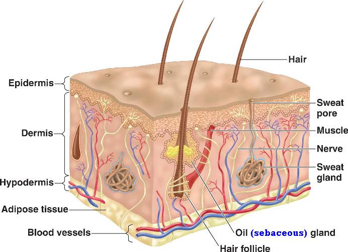
The integumentary system in our bodies is...wait for it.....our skin!
Yes the skin on your body is the integumentary system. It protects our bodies from the outside world. The air, the bugs in the world, and even the sun.
The epidermis is the top layer. It is completely dead skin. The skin is like a onion. It has many layers. Each layer rises and the dead skin is the top layer that is rubbed off. Interesting/ disturbing fact that I learned was you would have to wash your bed sheets everyday to get rid of the skin flakes that are rubbed off in your sleep.
Now the major layer in your integumentary system is the dermis. It holds major functions such as the the sweat glands and nerve endings. Also within this layer, are the capillaries. Another interesting fact is, when you lay in the sun (if fair skin you can see it the best) and you feel your skin heat up, it is your skin releasing the heat. The capillaries have expanded, making your skin seem red and is releasing the heat that is stored in your skin. It is acting as a refrigerator.
Now onto the fun part of this post. Skin diseases ....
Not all of them are dangerous but there are some that are life threatening. Such as... the bubonic plague (more commonly known as the black plague).
Which is caused by these little buggers
Which ride on these guys
Who stow away on boats and the flea jumps to people. Thus transferring the bacterial infection which caused painful swelling of the lymph nodes in your system and making them burst open from the overload of puss and blood. There are three types of the "plague". Not all of them have the swelling though so be careful.
Thursday, October 27, 2011
Friday, October 14, 2011
Tissue engineering review
Tissue Engineering
Summary:
The new development of bio genetic growing of tissue cells is now upon us. With the help of genetically altered rats, NASA, and the fore fathers of cell/tissue growth (Dr. Joseph Vacanti and Bob Langer). Tissue engineering is the basis for helping people who are needing to replace a organ or other features such as cardiac tissue, veins, cartilage, and skin. Thus it makes it easier for people who have a difficult time receiving help for a transplant. Way easier. The transplant actually works as well, even though it can be grown from a live specimen or even in NASA no-space container.
Weird example:

See! It's one of the genetically altered rats that is growing a ear for a person.... please don't shudder like I did.
Opinion:
*shudder fit*
Ok that is out of the way. See this is a new advancement from the past ( this all started in 1987). Now people don't have to be on the airing list for long anymore and people can also be treated for life threading illnesses like bone marrow transplants and cartilage transplants.
Summary:
The new development of bio genetic growing of tissue cells is now upon us. With the help of genetically altered rats, NASA, and the fore fathers of cell/tissue growth (Dr. Joseph Vacanti and Bob Langer). Tissue engineering is the basis for helping people who are needing to replace a organ or other features such as cardiac tissue, veins, cartilage, and skin. Thus it makes it easier for people who have a difficult time receiving help for a transplant. Way easier. The transplant actually works as well, even though it can be grown from a live specimen or even in NASA no-space container.
Weird example:

See! It's one of the genetically altered rats that is growing a ear for a person.... please don't shudder like I did.
Opinion:
*shudder fit*
Ok that is out of the way. See this is a new advancement from the past ( this all started in 1987). Now people don't have to be on the airing list for long anymore and people can also be treated for life threading illnesses like bone marrow transplants and cartilage transplants.
Tuesday, October 11, 2011
Histology
Histology: The study of the form of structures seen under the microscope. Also called microscopic anatomy, as opposed to gross anatomy which involves structures that can be observed with the naked eye. Traditionally, both gross anatomy and histology (microscopic anatomy) have been studied in the first year of medical school in the U.S. The word "anatomy" comes from the Greek ana- meaning up or through + tome meaning a cutting. Anatomy was once a "cutting up" because the structure of the body was originally learned through dissecting it, cutting it up. The word "histology" came from the Greek "histo-" meaning tissue + "logos", treatise. Histology was a treatise about the tissues of the body and the cells thereof.
Histology
I have studied cranial, epithelial,skeletal, and bone slides. Each were very different. In the lab we did, we were tought how to use a microscope and studied the afore mentioned slides under a 10x focus and then a 40x focus. we were told to draw and identify each slide. We did this and I shall post a good quality picture soon.
Epithelial:
This is just one example of epithelial tissue. It can also com in squamous, columnar, and pseudo-.
Cranial: is well from your head. It can also come in cuboidal, squamous, columnar, and pseudo-. There are no real good pictures I could find. Sorry.
Bone:
 Here are two really good pictures (one of which I stared at under a microscope which was extreamley amazing). This is bone tissue. You can tell it is by the circular pattern in the second picture.
This will also fall under the skeletal tissue catagory.
Anyways. I was also doing a lab with Cassy, and Jena. We learned how to use the microscope exceedingly well. Focusing was a cinch for us and we all took turns drawing what we saw. Unfortunately Jenna threw our drawings away because they could not be transferred to the computer well. This was a interesting lab, we had to guess at what we saw. Almost like a test to ourselves. And we had fun for this lab which made it worthwhile. Though we could not spell or pronounce any names we learned each slide slowly and it was worthwhile. I would like to thank Mr. Ludwig for that.
please forgive the spelling mistakes spell check broke on me
Histology
I have studied cranial, epithelial,skeletal, and bone slides. Each were very different. In the lab we did, we were tought how to use a microscope and studied the afore mentioned slides under a 10x focus and then a 40x focus. we were told to draw and identify each slide. We did this and I shall post a good quality picture soon.
Epithelial:
This is just one example of epithelial tissue. It can also com in squamous, columnar, and pseudo-.
Cranial: is well from your head. It can also come in cuboidal, squamous, columnar, and pseudo-. There are no real good pictures I could find. Sorry.
Bone:

This will also fall under the skeletal tissue catagory.
Anyways. I was also doing a lab with Cassy, and Jena. We learned how to use the microscope exceedingly well. Focusing was a cinch for us and we all took turns drawing what we saw. Unfortunately Jenna threw our drawings away because they could not be transferred to the computer well. This was a interesting lab, we had to guess at what we saw. Almost like a test to ourselves. And we had fun for this lab which made it worthwhile. Though we could not spell or pronounce any names we learned each slide slowly and it was worthwhile. I would like to thank Mr. Ludwig for that.
please forgive the spelling mistakes spell check broke on me
Subscribe to:
Comments (Atom)


